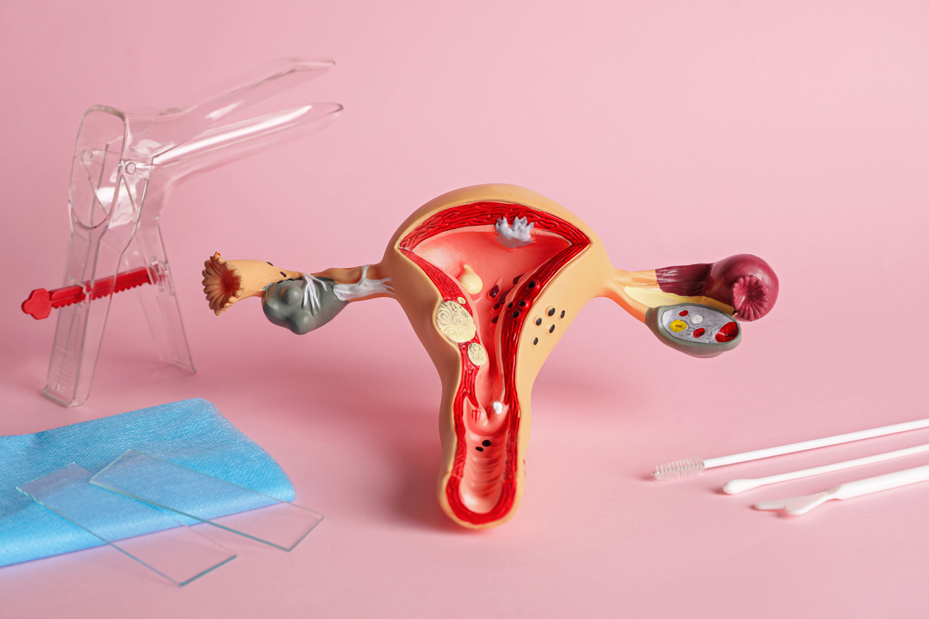
The uterus, an organ exclusive to women, is muscular, pear-shaped and located in the pelvis. During pregnancy, it’s able to stretch in order to accommodate the growing baby – then contracts and shrinks to its original size postpartum. Held in place by ligaments and muscles, these supporting structures weaken over time. When it’s unable to provide support, the uterus may sag or slip from its original position into the vagina causing a condition known as uterine prolapse.
Several factors may contribute to the weakening of the pelvic supports. Established risk factors include repeated vaginal births, aging, obesity, prior pelvic surgery, constipation, irritable bowel syndrome, genetic factors and neurologic injuries. There are also controversial risk factors which include episiotomy or intentional cutting of the perineum during childbirth, having macrosomic babies weighing more than 4500 grams at birth, heavy lifting and lower education.
Postmenopausal women are deemed to be at more at risk of developing this condition. Though age is a factor, women under 40 may still develop prolapse. Some mistakenly believe that this is only possible in women who delivered vaginally, and having a C-section is protective, but regardless of route of delivery or age, it’s really the growing uterus that creates stress on the supportive structures of the womb.
Women with milder cases may have no other obvious symptoms apart from the painless bulge. On the other hand, there are women suffering from moderate to severe prolapse –with the uterus slipping further out of place and exerting pressure on other pelvic organs, such as the bowels and the bladder. When this happens, the patient may manifest with discomfort or pain in the abdomen, pelvis and lower back. Pain during intercourse may also be observed and the patient may have unusual vaginal discharges.
Urinary symptoms may include urinary frequency or the need to urinate frequently, urinary incontinence or the involuntary loss of urine and urinary urgency or sudden urge to urinate.The patient may also suffer from recurrent urinary tract infections. In terms of bowel symptoms, constipation and feeling of fullness and pelvic heaviness are often present. These symptoms may be exacerbated during prolonged standing or walking due to the pull of gravity.
Apart from the symptoms reported by the patients, a pelvic examination is required to diagnose uterine prolapse. After a confirmed diagnosis, a management plan will be created depending on the severity of the prolapse — as well as the age, overall health status and fertility desires of the patient. Treatment may not always be necessary. Some cases are just managed by advocating healthy habits, exercise and avoidance of heavy lifting in order to avoid progression.
Non-surgical treatment options include Kegel exercises, which require tightening the pelvic muscles at will, as if trying to hold back urine. The muscles are held tight for a few seconds and then released and performed repeatedly for at least 10 times a day. Another non-surgical intervention uses a vaginal pessary –a rubber, plastic or silicone doughnut-shaped device measured to fit the lower part of the vagina to help prop-up the uterus and hold it in place.
A pharmacologic method in the form of estrogen creams showed improvement in vaginal tissue regeneration and strength. This is often used as an adjunct when employing conservative measures. Though relief is apparent in users, it doesn’t reverse the presence of a prolapse.

Surgical interventions include uterine suspension or vaginal hysterectomy. The former entails repair and re-attachment of pelvic ligaments that support the uterus. The latter is an operation which removes the uterus from the vagina which may or may not be accompanied by anterior and posterior colporrhaphy which strengthens the anterior and posterior wall of the vagina to prevent future prolapse of the remaining structures. This is ideal for severe cases of prolapse in patients who are post-menopause.
As effective as surgery is, it isn’t recommended for women who still want to conceive another child. Gravidity and childbirth can put an enormous strain on pelvic muscles, which can undo surgical repairs of the uterus.
How can one prevent development or progression of uterine prolapse? While there are no fail-proof preventions which can prevent all types of prolapse there are a few lifestyle tips that can help reduce one’s risk. Topmost on the list include: having or maintaining a healthy weight, doing regular exercises aimed at strengthening the pelvic floor muscles, eating nutritious meals, refraining from smoking and using proper lifting techniques.
Lucy* was a 46-year-old woman with four children, all delivered vaginally. She first noticed a small mass slightly peeking from her vagina while she was washing herself after defecation. The mass was non-tender but it was worrisome, because it was something unfamiliar. She immediately sought professional advice and was only prescribed with conservative management. Prompt consultation on her part prevented worsening of her condition and now, five years later, her prolapse remains at the same stage and doesn’t require surgery. Thorough counseling is key so that patients can make informed decisions and select the right treatment for themselves.
Accurate assessment, paired with appropriate and timely management of uterine prolapse can greatly impact a patient’s quality of life, including long-term physical and mental health effects.
*Name changed
Originally published in HealthToday Issue 1 2022
References:1. David Gershenson, Gretchen Lentz, Fidel Valea, Rogerio Lobo. Comprehensive Gynecology 8th Edition. Elsevier Publishing. May 2021. 2. Chen CJ, Thompson H. Uterine Prolapse. [Updated 2021 Nov 5]. In: StatPearls [Internet]. Treasure Island (FL): StatPearls Publishing; 2022 Jan-. Available from: https://www.ncbi.nlm.nih.gov/books/NBK564429/


